Case 2 180810-2 (18B0961)
Conference Coordinator: Dr Melissa Roy.
//
Five-year-old Quarter horse mare.
The owner noticed two 3 x 2 cm nodules on the left semimembranosus muscle, which the patient had been rubbing. They grew over the next three months, and one became ulcerated. Excisional biopsy with wide margins was performed.
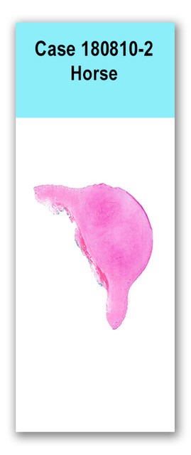
One small jar of formalin is received. The jar contained skin with two soft, smooth, round nodules measuring 1 cm in diameter and 2 cm in diameter. The larger nodule was focally ulcerated. On cut section the nodules were white, and smooth.
This slides has one section through a dermal mass, and the overlying haired skin. The mass is composed of an unencapsulated, moderately well demarcated, densely cellular mesenchymal neoplasm that expands the dermis and extends to the ulcerated overlying epithelium. The mass is composed of interlacing streams and whorls within a variably dense basophilic to collagenous fibrovascular stroma. Cells are spindloid and have indistinct cell borders and scant eosinophilic to amphophilic cytoplasm. Nuclei are ovoid to round, with finely stippled chromatin and one variably distinct nucleolus. There is marked anisocytosis and anisokaryosis and 6 mitotic figures within 10 400x fields. At the dermo-epidermal junction, neoplastic cells are arranged perpendicularly to the surface. Extending from the moderately well-demarcated mass, there are similar cells extending along the dermis, weaving between collagen bundles, and to the margins. The ulcerated region of epidermis is necrotic and contains degenerate neutrophils, fibrin, hemorrhage, and basophilic karyorrhectic debris. Intact epithelium has multifocal rete pegs extending into the dermis.
N/A
Haired skin: Sarcoid
The majority of the neoplastic cells are within a relatively well-demarcated nodule from which the lateral margins are clear; however, the dermal collagen extending from that nodule has a small amount of similar mesenchymal cells weaving between collagen bundles. We cannot say with certainly whether these are part of the neoplastic population. If more definitive margins are desired, identification of bovine papilloma virus by in situ hybridization can be performed to identify cells of interest. Without this, interpretation of calculated margins should be done with caution.
Case contributor:
Drs. Melissa Roy, Chuck Mohr, and Verena Affolter. Conference presenters: Drs. Melissa Roy and Kevin Keel