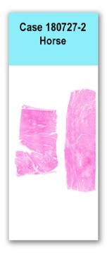
Case 2 180727-2 (17N3045)
Conference Coordinator: Dr Elizabeth Rose.
//
thirteen-year-old, quarterhorse mare
The patient presented with a two-day history of deterioration with minimal response to banamine. Physical exam revealed tachycardia, hyperemic mucous membranes and ataxia with intermittent episodes of pyrexia and reduced fecal output. Clinicians were suspicious for colitis-induced endotoxemia and euthanized the patient due to poor prognosis and lack of response to treatment.
The lungs failed to collapse and oozed red, clear, watery fluid. There were approximately 5 to 10 L of red to yellow, clear, watery fluid within the thoracic cavity. Diffusely spanning the epicardium overlying the right auricle and multifocally extending along the coronary groove to the epicardium overlying the left and right ventricles were dozens of irregularly-marginated to linear, smooth, flat, mottled bright red/dark red/dull, dark purple discolorations. On section, the discolorations spanned the myocardium of the right auricle and extended approximately 3 mm deep into the myocardium of the right and left ventricles. The epicardium overlying the right ventricle was multifocally pale tan and roughened; the pale discoloration extended up to 1cm deep into the myocardium. Across the entire endocardium were 10 to 15, multifocal to coalescing, dark red to dull purple, flat discolorations that measured up to 2 cm in diameter and extended up to 2 mm deep into the underlying myocardium. The endocardium of the left ventricle was most severely affected.
Along the distal half of the esophagus were dozens of linear and circular ulcers that measured up to 8 cm long and 0.5 cm in diameter. The ulcer margins were flat and tattered (acute). The central regions were red to brown (muscularis subjacent to the mucosal defects was completely exposed). Affecting approximately 30% of the nonglandular portion of the stomach, there were punctate to 2 mm in diameter ulcers, covered with soft brown gelatinous fluid (clots).Two sections of ventricular myocardium are examined in which there are multifocal to coalescing, randomly-scattered regions of cardiomyocyte necrosis characterized by hypereosinophilia, pyknotic nuclei, contraction bands and fragmentation of the cytoplasm. Occasionally, affected myocytes have cytoplasmic, deeply basophilic stippling (interpreted as dystrophic mineralization). These regions are surrounded by abundant lymphocytes, plasma cells, histiocytes and extravasated red blood cells. A similar inflammatory population extends into the epicardium and endocardium, which are expanded by clear space (edema) and lakes of extravasated red blood cells.
N/A
Ancillary Diagnostics: Oleandrin was identified in the stomach contents. Trace amounts of oleandrin were also evident in the cecal contents.Heart: Severe multifocal myocardial degeneration and necrosis with mild multifocal lymphoplasmacyti histiocytic neutrophilic myositis and moderate hemorrhage
Toxicological analysis of the stomach and cecal contents confirmed oleandrin toxicosis as the cause of the patient’s acute demise and myocardial lesions. Pink oleander (Nerium oleander) is a popular ornate shrub and all parts of the plant are highly toxic. Oleandrin is a cardioactive glycoside, the primary targets of which are the heart and gastrointestinal tract. More rarely, neurologic signs and renal tubular degeneration have also been reported. Through interference with the normal calcium channel and eventual intracellular accumulation of calcium, oleandrin exerts a positive inotropic effect, meaning it increases the strength of muscular contractions. Multifocal endocardial and epicardial hemorrhages, especially of the right auricle, are common gross lesions. The lesions in this case were acute and monophasic, which is expected with an acute toxicity.
Other cardiotoxic plants that should be considered include, foxglove, mountain laurel, azaleas, rhododenrons, yew, gossypol, locoweed. Ionophores (monensin and salinomycin) can also cause myocardial necrosis. The contributors of the case also initially considered cantharidin due to the severe ulceration of the esophagus and stomach. Without any history of the case, cardiotoxicity would be difficult to definitively distinguish from ischemic myocardial necrosis, which can be caused by disseminated intravascular coagulopathy, vitamin E/selenium deficiency and inflammatory diseases. Vitamin E and selenium toxicological analysis of this patient’s liver was within normal limits.1. Sharma P, Choudhary AS, Parashar P, Sharma MC, Dobhal MP. Chemical constituents of plants from the genus Nerium. Chem Biodivers. 2010;7(5):1198-207
2. Omidi A, Razavizadeh AT, Movassaghi AR, Aslani MR. Experimental oleander (Nerium oleander) intoxication in broiler chickens (Gallus gallus). Hum Exp Toxicol. 2011 May 16. Case contributor: Dr. Liz Rose and Dr. Patti Pesavento
