Case 1 180720-1 (T17026608-7)
Conference Coordinator: Dr Sarah Stevens.
//
Thirty-day-old female Holstein calf.
Euthanized due to Salmonella Dublin septicemia and chronic bronchopneumonia (Trueperella pyogenes and Mycoplasma bovis cultured).
Multifocal to coalescing areas of scalloping/ulceration were observed within the oral cavity following the mandibular molars. These areas were overlain by a layer of thick white material that can be scraped off. Similar areas of ulceration are observed on the tongue and variably appear targetoid.
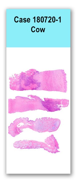
This slide has four sections of tissue, two of oral mucosa with underlying salivary glandular tissue and two sections of tongue. The sections of tongue have variable, moderate to severe, mucosal epithelial hyperplasia with moderate, parakeratotic hyperkeratosis. Multifocal single cell apoptosis/necrosis of superficial epithelial cells is intermixed with cells undergoing ballooning degeneration. Very rarely intracellular (unclear on intracytoplasmic versus -nuclear) discrete, dark eosinophilic inclusions are identified. The basal layers of the mucosal epithelium are moderately jumbled forming rounded, moderately thick rete pegs. The immediate submucosa is mildly edematous with moderate numbers of scattered lymphocytes, plasma cells and neutrophils. Similar inflammatory cells trickle down into the deeper connective tissue and skeletal muscle, which has multifocal large regions of necrosis admixed with degenerate to fragmented inflammatory cells and abundant colonies of bacteria (apparently rod-shaped).
The two sections of oral mucosa and underlying submucosa with salivary gland are similarly inflamed with generalized, moderate, mucosal epithelial hyperplasia with one section also affected by regional necrosis with similar bacterial infiltration.Gram stain: There are abundant bacterial colonies, which predominantly appear rod-shaped and gram-negative.
Tongue: Severe, subacute, multifocal focal proliferative and necrotizing glossitis with abundant intralesional bacteria Gingiva: Severe, subacute, regionally extensive necrotizing and proliferative gingivitis
The gross, histologic and PCR findings are consistent with a diagnosis of bovine papular stomatitis. In addition, the necrotizing component of the stomatitis was attributed to the abundant intralesional bacteria, which was consistent with Fusobacterium necrophorum based on histology, gram stain results and bacterial culture.
Conference participants discussed whether there was a relationship between these two entities in this case or if it was equally as possible that these processes occurred independently of one another as F. necrophorum isn’t particularly associated with an initial viral infection. In this case, the relationship between the two processes can’t be definitively determined but either scenario is likely possible. The individual who contributed this case was somewhat interested in the bacterial culture results as the gross appearance of the lesions almost appeared “fuzzy” and therefore, there was initial suspicion for a fungal infection such as Candida sp.N/A