Case 4 180615 (18N1147)
Conference Coordinator: Devinn Sinnott.
//
20-year-old, male, intact, golden lion tamarin (Leontopithecus rosalia)
The patient had a two-day-long history of anorexia, weakness, lethargy, and staring blankly. No physical exam or blood work was performed, and humane euthanasia was elected.
One kidney had an approximately 5-mm-diameter, irregularly shaped, well demarcated, soft, pale-tan nodule within the cortex and medulla. A second, similarly sized, irregularly shaped, well demarcated, soft, mottled tan and dark red mass was also present in the cortex and medulla of the same kidney.
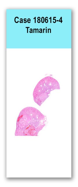
Two sections of kidney are examined in which the cortex is compressed by three large, nodular, well demarcated, partially encapsulated, densely cellular masses. The masses are composed of polygonal cells arranged in variably sized islands and papillary projections supported by thin stalks of fibrovascular tissue. Cells have distinct cell borders and abundant wispy, pale eosinophilic to clear cytoplasm. Nuclei are round with finely stippled chromatin and a variably distinct nucleolus. Anisocytosis and anisokaryosis are mild, and three mitotic figures are seen in ten 400x fields. Large lakes of hemorrhage admixed with eosinophilic, fibrillar material (fibrin), sloughed neoplastic cells, karyorrhectic debris, and macrophages with intracytoplasmic, finely granular, gold-brown pigment are scattered throughout the masses. The renal tubules adjacent to the mass are frequently dilated and lined by variably attenuated epithelium. These tubules often contain brightly eosinophilic, homogenous, intraluminal material, and are surrounded by moderate numbers of lymphocytes and plasma cells.
N/A
Kidney: Clear-cell renal carcinoma with intra-tumoral hemorrhage Kidney: Moderate, chronic, multifocal, lymphoplasmacytic, interstitial nephritis with tubular dilatation, eosinophilic tubular casts and epithelial attenuation (thyroidization)
Primary renal tumors are rare in callitrichids despite their common use as laboratory animals. A primary renal adenoma has been reported in a cotton-topped tamarin, as well as a nephroblastoma. In domestic species, renal carcinomas are also rare; however, they are the most common primary renal tumor in dogs, cats, and horses. Histologic subtypes include solid, tubular, papillary, and cystic, and these are further categorized cytologically as chromophobic, eosinophilic, and clear cell variants. Clear cell variants are rare in dogs (9% of renal carcinomas) but account for the vast majority of human renal carcinomas (75%). This variant is derived from the proximal convoluted tubular epithelium, and often grows in a solid pattern. The clear cytoplasm is derived from abundant intracytoplasmic glycogen and lipid storage.
Some reviewers of this slide commented that the pattern of thyroidization is thought to be associated with pyelonephritis.
Brack M. Renal papillary adenoma in a cotton-topped tamarin (Saguinus Oedipus). Laboratory Animals 1985; 19: 132-133.
Meuten DJ (ed.). Tumors in domestic animals. 5th edition. Ames, IA: Wiley Blackwell; 2017. p. 638-645.