Case 2 180615-2 (18N1194)
Conference Coordinator: Devinn Sinnott.
//
3-year-old, female chicken
The patient was attacked by a dog, and several days later the owner noticed that she was emaciated and frequently regurgitated. The referring veterinarian performed an ingluvotomy. At a recheck examination several days later the incision had dehisced and the coelomic cavity was distended. Gastric lavage was recommended, but during anesthesia the patient died.
The ovary was effaced by a 5 x 5 x 3 cm, irregularly shaped, multinodular, firm, pink to pale tan mass. Similar, 3- to 8-mm-diameter nodules were randomly scattered throughout the mesentery and along serosal surfaces of the coelomic viscera. The mass slightly displaced the ventriculus ventrocaudally.
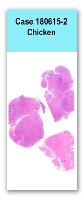
Two sections of ovary and one section of ventriculus are examined. The ovary is expanded by a large, multinodular, poorly demarcated, unencapsulated, infiltrative, densely cellular mass. The mass consists of polygonal cells arranged in tubules and variably sized islands surrounded by a dense collagenous stroma. Tubules occasionally contain pale eosinophilic, wispy material. Cells have variably distinct cell borders and a small to moderate amount of eosinophilic, finely vesiculated cytoplasm. Nuclei are round and euchromatic with 1 to 2 nucleoli. Anisocytosis and anisokaryosis are mild, and three mitotic figures are seen in ten 400x fields. Three similarly appearing, encapsulated nodules are present on the serosal surface of the ventriculus.
N/A
Ovary: ovarian carcinoma with intracoelomic serosal implantation (carcinomatosis)
Ovarian carcinomas are relatively common neoplasms in domestic chickens, with an incidence of up to 24% in laying hens over two years of age. As in humans, these tumors are believed to occur due to DNA damage to the ovarian surface epithelium secondary to repeated rupture of the epithelium during ovulation. Ovarian carcinomas in chickens express similar molecular markers to human ovarian carcinomas, including cytokeratin, epidermal growth factor receptor (EGFR), Tag 72, proliferating cell nuclear antigen (PCNA), TGF-a, CA125, E-cadherin, and p53. Because of these similarities in pathogenesis and molecular behavior, laying hens are an important animal model for human ovarian carcinoma studies. Intracavitary implantation can occur due to rupture of cysts within the tumor or invasion through the ovarian capsule, often leading to ascites or abdominal distension due to blockage of lymphatics by neoplastic cells.
Fredrickson TN. Ovarian tumors of the hen. Environmental Health Perspectives 1987; 73: 35-51.
Hawkridge AM. The chicken model of spontaneous ovarian cancer. Proteomics Clin. Appl. 2014; 8: 689-699.
Barua A, Bitterman P, Abramowicz JS, Dirks AL, Bahr JM, Hales DB, Bradaric MJ, Edassery SL, Rotmensch J, Luborsky JL. Histopathology of ovarian tumors in laying hens, a preclinical model of human ovarian cancer. Int. J. Gynecol. Cancer 2009; 19(4): 531-539.
Meuten DJ (ed.). Tumors in domestic animals. 5th edition. Ames, IA: Wiley Blackwell; 2017. p. 690-694.