Case 3 180511 (18N0640)
Conference Coordinator: Dr Elizabeth Rose.
//
Twenty-four-year-old warmblood mare
The patient was presented for a two-month-long history of severe unilateral hindlimb lameness. Three days earlier, a referring veterinarian drained an abscess in the toe region, however the patient did not improve despite a full dose of banamine and was referred to UC Davis. The patient was euthanized due to poor prognosis for future soundness.
The cranial third of the sole of the left hindlimb is severely eroded or ulcerated, exposing the lamina and the third phalanx (P3). There is a similar, approximately 1-cm-diameter, focus of ulceration in the frog of the hoof. The remainder of the sole and frog is almost completely separated from the adjacent lamina, and is attached only at the caudal third of the hoof. The distal half of P3 is severely cavitated, with an irregularly shaped, 3- to 5-mm-wide bony rim remaining. The adjacent lamina is homogenously soft, friable and mottled pale pink to bright red. The lateral margins of the right hindlimb sole are eroded to ulcerated, in a manner that is similar to but less severely affected than that of the left forelimb. The junction of the cranial-most tip of the sole and the distal hoof wall contains a 2 cm in diameter, full-thickness ulceration.
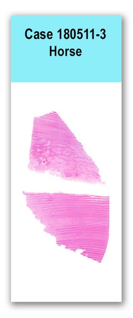
Two sections of primary and secondary laminae are examined. Dermal and epidermal lamella are multifocally effaced by large, irregularly-shaped islands and bands of dense collagen and multifocal spicules of mineralized bone. Collagen islands are surrounded by dense fibrovascular tissue that is heavily infiltrated with neutrophils, lymphocytes, plasma cells and histiocytes. The secondary dermal lamellae are diffusely blunted to absent. The secondary dermal lamellae are occasionally, multifocally separated from the secondary epidermal lamellae.
No special stains.
Moderate to severe multifocal chronic neutrophilic lymphoplasmacytic laminitis with epidermal laminar degeneration, necrosis and hyperkeratosis.
This case was chosen to highlight the histologic architecture of lamina and defining features of laminitis. The horse hoof is attached to the third phalanx by the interdigitation of the secondary epidermal lamina and the secondary dermal lamina. Laminitis occurs when the epidermal and dermal lamina separate, the pathogenesis of which is poorly understood. In early stages of laminitis, the normally club-shaped ends of the secondary epidermal lamina become elongated and attenuated at the tips. The epidermal cells detach from the underlying basement membrane, occasionally forming teat-shaped bubbles. The detachment is best appreciated with a periodic acid-schiff stain. Lesions may progress to full retraction of the basement membrane and the secondary dermal lamina from the secondary epidermal lamina. As the epidermal lamina becomes further detached from its blood supply, subsequent ischemia may occur. Clinically-appreciated bounding pulses are consequent to increased resistance to blood flow within the retracted capillaries. In the most severe cases of laminitis, there is a completely loss of distinction between primary and secondary epidermal lamina.
Pollitt CC. Basement membrane pathology: a feature of acute equine laminitis. Equine Vet J. 1996;28:38-46.
Ginn PE, Mansell JEKL, Rakich PM. Skin and appendages. In: Maxie MG, ed. Jubb, Kennedy, and Palmers Pathology of Domestic Animals. 5th ed. Toronto, CA: Saunders Elsevier; 2007:553-781.
Case contributor: Drs. Rose and Woolard
Case presenter: Drs. Rose