Case 4 180504 (18B0435)
Conference Coordinator: Devinn Sinnott
//
An approximately six-month-old, male, castrated domestic short-hair cat
The patient was referred to an ophthalmology service for bilateral enucleation after a referring veterinarian diagnosed him with bilateral blindness, congenital glaucoma, buphthalmia, anterior uveitis, and lens subluxation.
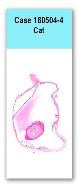
A 2.5-cm-diameter globe was received in which the cornea was diffusely cloudy. On cut section the posterior chamber was dilated, but the eye was otherwise unremarkable.
A parasagittal section of an eye is examined. Proteineaceous exudate is present in the chambers; the uvea is heavily infiltrated by plasma cells; and the ciliary cleft is collapsed. The cornea is multifocally infiltrated by neutrophils. A thin margin of immaure blood vessels extends through the corneal stroma from the limbus, and the stroma has decreased clefting (edema). The corneal epithelium is focally ulcerated with disorganization of the detached epithelium. It is variably attenuated, and often has vesicles of protein-rich fluid within the remaining epithelium. The endothelial aspect of the cornea has multiple adherent aggregates of plasma cells, neutrophils, and macrophages admixed with fibrin (keratic precipitates). The posterior aspect of the lens capsule is ruptured and curled, and the cortical and subcapsular lens fibers are multifocally swollen, liquified, and disorganized. A coagulum of plasma cells and fewer neutrophils and macrophages admixed with fibrin (thrombus) extrudes from the posterior chamber, through the sclera, and onto the episcleral surface. The retina is mildly infiltrated by lymphocytes and plasma cells, has a mildly decreased number of ganglion cells, and is focally detached with mild subretinal hemorrhage. The optic nerve is severely cupped with swollen axon sheaths and axon loss, gliosis, disorganization, and leakage of vitreous within the nerve.
Immunohistochemistry for feline coronavirus showed scattered positively staining cells (consistent with macrophages) within the keratic precipitates and freely floating in the anterior chamber of the eye
1. Severe, chronic, diffuse, plasmacytic panuveitis with keratic precipitates 2. Mild, chronic, diffuse, lymphoplasmacytic retinitis with focal detachment 3. Chronic secondary glaucoma 4. Cortical and subcapsular cataract with suspected lens capsule rupture 5. Severe, chronic, multifocal, suppurative keratitis with limbal neovascularization and corneal edema
The prevalence of ocular lesions in feline infectious peritonitis (FIP) is unknown, but has been clinically estimated at 10-50%. However, most cats that die of FIP have ocular involvement as evidenced by coagulation of the aqueous by acidic fixative, indicating an elevated protein content. The manifestation of FIP in the eye varies widely, and can be quite subtle. The inflammatory infiltrate is typically composed of equal numbers of neutrophils, macrophages, lymphocytes, and plasma cells, but can be purely histiocytic in some cases. The predominantly plasmacytic nature of the inflammation in this case is unusual, but aggregates of plasma cell-rich inflammation is very useful in the diagnosis of FIP. Keratic precipitates, as seen in this case, are a common histologic feature of ocular FIP.
Maxie G, editor. Jubb, Kennedy, and Palmer's Pathology of Domestic Animals, 5th edition. New York: Elsevier Saunders; 2007. Volume 1, p. 505.
Dubielzig RR, Ketring K, McLellan GJ, Albert DM. Veterinary Ocular Pathology: A Comparative Review. Edinburgh: Elsevier Saunders; 2010. p. 268-270.
Case contributor:
Dr. Chris Reilly was consulted for this case.
Conference presenters:
Dr. Devinn Sinnott and Dr. Brian Murphy