Case 3 180504 (18N0575)
Conference Coordinator: Devinn Sinnott
//
Three-year-old, female, spayed Yorkshire terrier
In February 2018, the patient was presented to a referring veterinarian for progressive dyspnea, and was diagnosed with an interstitial pattern on radiographs and unremarkable blood work. Two days later, radiographs were repeated and the patient was diagnosed with a pneumomediastinum. She was hospitalized for ten days, and she was treated with antibiotics and steroids. She improved slightly and was sent home for a week before begin presented to the referring veterinarian again. The pneumomediastinum was still present; a cervical exploratory surgery was performed to look for a possible tracheal rupture but none was found. The patient presented to an emergency service in March 2018, in severe respiratory distress. She was hospitalized in ICU overnight. Due to concerns for quality of life, the owners elected humane euthanasia.
Negative pressure was present in the thoracic cavity. The mediastinum was expanded by air from the thoracic inlet to the diaphragm. The lungs were soft and mottled pink and dark red, and the cranioventral lung lobes were dark red and slightly firmer than the surrounding parenchyma. The pleural surface of all lobes had approximately 25-50, irregularly shaped, well demarcated, smooth, gray discolorations ranging in diameter from approximately 2 to 5 mm that did not extend into the subjacent parenchyma. The left cranial and left caudal lobes each had a round, well demarcated, 1-cm-diameter, smooth, soft, pink discoloration of the pleural surface, and the underlying parenchyma was soft and dark red.
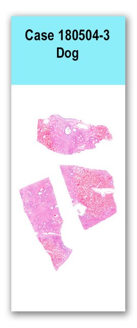
Three sections of lung are examined in which the interstitium and alveoli in multiple foci are expanded by a poorly demarcated, unencapsulated infiltrate of pleomorphic cells that form irregular tubules and nests and are occasionally individualized. These cells have variably distinct cell borders, moderate to large amounts of finely vesiculated, eosinophilic cytoplasm, and one to two round to ovoid nuclei with finely stippled chromatin and one to two distinct, basophilic nucleoli. Anisocytosis and anisokaryosis are marked, and one mitotic figure is seen in ten 400x fields. Individual cells are frequently necrotic. The interstitium is additionally expanded by a marked amount of fibrosis, fibrin, and a small number of lymphocytes and plasma cells. Alveoli are often filled with large, pleomorphic, foamy macrophages, erythrocytes, lymphocytes, and plasma cells.
Immunohistochemistry with pancytokeratin highlighted scattered islands of highly pleomorphic, neoplastic epithelial cells. Immunohistochemistry with CD18 showed abundant large, atypical histiocytes within the interstitium and alveoli.
Lungs: Pulmonary carcinoma (lepidic subtype)
Lungs: Moderate, chronic, multifocal interstitial fibrosis with interstitial and alveolar histiocytosisBased on the marked pleomorphism and poor demarcation of the clusters of pancytokeratin positive cells, a pulmonary carcinoma was diagnosed. The multifocal and infiltrative nature of the carcinoma as well as the young age of the patient is unusual, and prompted the idea of a possible inhaled carcinogen. However, based on the detailed history provided by the owner, toxin exposure was unlikely for this patient. A possible differential diagnosis is severe bronchioloalveolar hyperplasia and dysplasia secondary to chronic inflammation or toxin exposure. Abundant pleomorphic histiocytes are also present within alveoli and the interstitium, but these are presumed to be in response to the neoplasm.
Case contributor:
This case was received on necropsy service by Dr. Devinn Sinnott and Dr. Verena Affolter.Conference presenters: Dr. Devinn Sinnott and Dr. Brian Murphy