Case 2 180504-2 (18N0532)
Conference Coordinator: Devinn Sinnott
//
Thirteen-year-old, female, intact Canada lynx (Lynx canadensis)
The patient was rescued from a small zoo in 2012. She had a bout of leptospirosis in May 2016, from which she recovered. In March 2018, she suddenly became lethargic, became anorexia, and developed mild tachypnea. She was found dead the next morning. The property had animals that were previously diagnosed with Franciscella tularensis and Yersinia pseudotuberculosis several years prior. The property does not use pesticides, insecticides, or rodenticides, but is near agricultural lands.
The adipose tissue within the cranial mediastinum had multifocal to coalescing areas of dark red discoloration that extended within the tissue. The parietal pleura along the ribs had greater than 100, pinpoint, dark red, smooth discolorations. The lungs were diffusely atelectatic, soft, and dark red.
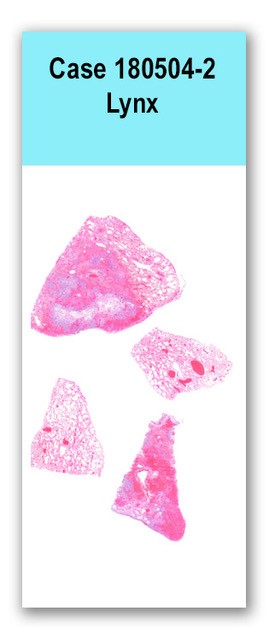
Four sections of lung are examined in which two sections have alveoli filled with erythrocytes, fibrin, large numbers of neutrophils, and smaller numbers of histiocytes, lymphocytes, and plasma cells with intra- and extracellular clouds of coccobacilli. Alveolar septae are often necrotic and expanded by fibrin. Vessels within necrotic and hemorrhagic regions of the parenchyma often contain thrombi which occlude the lumen and obscure the intimal layer. The muscular walls of these vessels are expanded by brightly eosinophilic, fibrillary material (fibrinoid necrosis). Some bronchioles are necrotic with loss of the epithelium and filling of the lumen with hemorrhage, fibrin, and a similar inflammatory population. The pleura is multifocally necrotic and covered in fibrin. In less affected regions of the parenchyma, the alveoli contain fewer inflammatory cells, fibrin, and coccobacilli
A Gram stain confirmed the presence of large clouds of gram-negative rods. Culture of the lung yielded hemolytic Escherichia coli.
Lungs: Severe, acute, multifocal, neutrophilic and necrohemorrhagic bronchopneumonia with intra-alveolar fibrin, fibrinoid vascular necrosis and thrombosis, and intralesional coccobacilli
Sudden death can be attributed to effacement of large portions of the lungs by severe inflammation and hemorrhage. The hemolytic E. coli cultured from the lung tissue is consistent with the Gram stain results which showed large clouds of gram-negative, rod-shaped bacteria. The source of the E. coli is uncertain. No other tissues examined showed signs of similar inflammation or sepsis. Large numbers of bacteria were seen in the colon; however, there was no corresponding inflammation or compromise of the intestinal wall that may have led to bacterial translocation and hematogenous spread to the lungs. An inhalational route of infection is presumed.
Escherichia coli has been reported as a cause of acute, necrotizing and hemorrhagic pneumonia in puppies, kittens, adult cats, and a tiger cub. In these cases, infection was thought to have occurred due to immunocompromise from young age or co-infection. In the case of the lynx, no other significant comorbidities were noted histologically. In the previously reported cases, certain strains of E. coli were also associated with infection. Further typing of the E. coli isolated from the lynx lung was not pursued. A recent study using a rodent model of ventilator-associated E. coli pneumonia in humans suggests that the genes involved in lung pathogenicity differ from those of other extraintestinal infections.Handt LK, Stoffregen DA, Prescott JS, Pouch WJ, Ngai DTW, Anderson CA, Gatto NT, DebRoy C, Fairbrother JM, Motzel SL, Klein HJ. Clinical and microbiologic characterization of hemorrhagic pneumonia due to extraintestinal pathogenic Escherichia coli in four young dogs. Comparative Medicine. 2003. 53(6): 663-670.
Breitschwerdt EB, DebRoy C, Mexas AM, Brown TT, Remick AK. Isolation of necrotixigenic Escherichia coli from a dog with hemorrhagic pneumonia. Journal of the American Veterinary Medical Association. 2005. 226(12): 2016-2019.
Highland MA, Byrne BA, DebRoy C, Samitz EM, Peterson TS, Oslund KL. Extraintestinal pathogenic Escherichia coli-induced pneumonia in three kittens and fecal prevalence in a clinically healthy cohort population. Journal of Veterinary Diagnostic Investigation. 2009. 21: 609-615.
4. Sura R, Van Kruiningen HJ, DebRoy C, Hinckley LS, Greenberg KJ, Gordon Z, French RA. Extraintestinal pathogenic Escherichia coli-induced acute necrotizing pneumonia in cats. Zoonoses and Public Health. 2007. 54: 307-313.
Carvallo FR, DebRoy C, Baeza E, Hinckley L, Gilbert K, Choi SJ, Risatti G, Smyth JA. Necrotizing pneumonia and pleuritis associated with extraintestinal pathogenic Escherichia coli in a tiger (Panthera tigris) cub. Journal of Veterinary Diagnostic Investigation. 2010. 22:136-140.
Phillips-Houlbracq M, Ricard JD, Foucrier A, Yoder-Himes D, Gaudry S, Bex J, Messika J, Margetis D, Chatel J, Dobrindt U, Denamur E, Roux D. Pathophysiology of Escherichia coli pneumonia: Respective contributions of pathogenicity islands to virulence. International Journal of Medical Microbiology. 2018. 308(2): 290-296.
Case contributor:
This case was received on necropsy service by Dr. Devinn Sinnott and Dr. Verena Affolter.
Conference presenters:
Dr. Devinn Sinnott and Dr. Brian Murphy