Case 4 180427 (18N0451)
Conference Coordinator: Wesley Siniard.
//
9-year-old, intact female pot belly pig
This animal had a several month history of hyporexia and straining to defecate and urinate. Radiographs revealed a large soft tissue mass within the abdominal cavity. An exploratory laparotomy was performed, which confirmed the large abdominal mass, which was believed to be originating from the uterus. Numerous adhesions were noted between the mass and other abdominal organs. Due to poor prognosis, humane euthanasia was elected.
Approximately two liters of dark red, watery fluid was in the abdominal cavity. The serosal surfaces of all abdominal organs as well as the abdominal body wall were covered with large amounts of red to tan, stringy, friable material (fibrin). A 16.2 kg, 38 x 40 x 23 cm, firm mass which was mottled pink to tan was originating from the uterine body wall. Both uterine horns extended to and blended into the mass. On cut surface, the mass was multilocular and mottled tan to brown to yellow. A 18 x 18 x 5 cm, firm, pale tan, multinodular mass was in the left mesometrium. On cut surface, it was diffusely pale tan. The mucosal surface of the uterus had 500 to 600, multifocal, fluid filled cysts which measured from 0.3 cm to 2 cm in diameter. Fluid within the cysts was pale yellow and watery. The serosal surface of all intestines was mottled dark red to yellow to brown, and was overlain by a layer of fibrin.
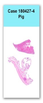
One section of uterus is examined, in which a small, well demarcated, unencapsulated, expansile, multinodular, densely cellular mass is arising from the myometrium. Mass is composed of spindle cells arranged in short intersecting streams and bundles within a fine fibrovascular stroma. Cells have variably distinct cell borders, a small amount of pale eosinophilic, finely fibrillar cytoplasm, and a single elongate nucleus with finely stippled chromatin and occasionally a prominent basophilic nucleolus. Anisocytosis and anisokaryosis are mild, and 1 mitotic figure in 10 hpf. Endometrium is expanded by numerous markedly dilated glands lined by 1-3 layers of cuboidal to columnar, infrequently ciliated epithelial cells and contain variable amounts of pale eosinophilic, foamy material or brightly eosinophilic, homogeneous material. Submucosal stroma surrounding these glands is mildly infiltrated with small numbers of lymphocytes and plasma cells.
N/A
Uterus: Cystic endometrial hyperplasia
Uterus: Focal vascular metastatic leiomyosarcoma
Pigs such as pot belly pigs live longer than those used for production, and therefore are predisposed to development of reproductive tract lesions. In a recent article assessing reproductive lesions in pet pigs (Ilha MRS, 2010), cystic endometrial hyperplasia was determined to be the most prevalent gross and histologic change noted in the pigs examined with uterine lesions. In this paper, 44% of the pigs examined also had concurrent smooth muscle tumors, suggesting that there may be an association between the two entities. In this case, two large leiomyosarcomas were noted in the uterus, with one originating from the uterine body, and one originating from the mesometrium of the uterine horn. The metastatic lesion present in the section submitted, is likely a metastasis from one of these primary tumors; however, metastasis of uterine leiomyoma / leiomyosarcomas is rare.
Ilha MRS, Newman SJ, Van Amstel S, Fecteau KA, Rohrbach BW. Uterine Lesions in 32 Female Miniature Pet Pigs. Veterinary Pathology. 2010. 47(6) 1071-1075.