Case 3 180525 (18B0984)
Conference Coordinator: Dr Melissa Roy.
//
Seven-month-old, female, intact golden mixed breed dog (golden retriever X poodle)
One month prior to presentation, the owners noted that the patient combat crawling; and that her left pelvic limb was dragging and causing bleeding from a nail on that limb. An MRI revealed an intradural, extramedullary mass at the level of the T12 spinal cord, and she was prescribed prednisone. One month later, with some clinical improvement, the patient presented to the VMTH for further evaluation. At that time, the patient underwent a right hemilaminectomy and mass debulk.
The mass was tightly adhered to the spinal cord and only able to be 90% excised. There was significant compression of cord and secondary changes to cord (edema, softness, abnormal blood vessels).
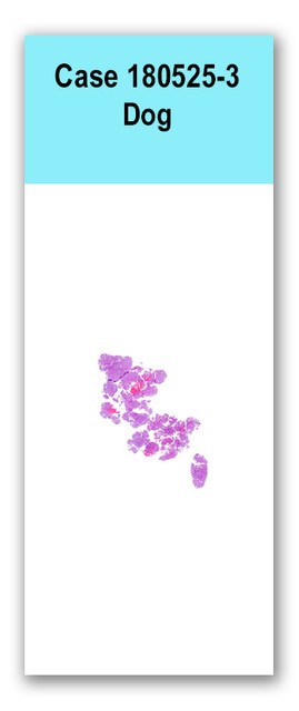
This slide has fragmented pieces from a section of intradural spinal cord mass. The tissues examined consist entirely of a densely cellular, unencapsulated mass which extends to all borders, such that interpretation of the relationship to surrounding tissues is not possible. Neoplastic cells are arranged in a variety of patterns. Some cells are arranged in tubules lined by cuboidal to columnar cells with variably distinct cell borders, a small amount of eosinophilic cytoplasm, and one central irregularly round to ovoid nucleus with finely stippled chromatin. Tubules occasionally have densely cellular tufts projecting into the lumen (primitive glomeruli). The rest of the neoplastic cells surrounding tubules are arranged in sheets, streams, or vague whorls within a fine fibrovascular stroma. These cells have a small amount of eosinophilic cytoplasm, indistinct cell borders, and round to ovoid nucleus with vesiculated chromatin and one variably distinct nucleolus. Occasionally, cells are clumped into nests composed of polygonal cells with indistinct cell borders, scant eosinophilic cytoplasm and one nucleus with dense chromatin. Anisocytosis and anisokaryosis are moderate, and there are 23 per ten 400X fields, including occasional bizarre mitotic figure. Multifocal regions of hemorrhage are scattered throughout the mass.
No special stains.
Thoracolumbar spinal tumor of young dogs (spinal nephroblastoma)
The combination of anatomical location and histomorphology are consistent with a spinal nephroblastoma, also termed thoracolumbar spinal tumor of young dogs. Although present in the spinal cord, this is an embryonal tumor that arises from non-neuroepithelial cells, likely remnant renal precursors entrapped in the spinal cord during development. They are most often intradural and extramedullary. The best immunohistochemical assay for this type of tumor is Wilms' tumor 1 (WT1), which is not available in our histology laboratory. These tumors are almost invariably located between the tenth thoracic vertebra (T10) and the second lumbar vertebra (L2). The often occur in large-breed dogs, and extra-spinal involvement is not reported.
We thank Dr. Kevin Woolard for contributing this case.