Case 4 180810 (18B1630)
Conference Coordinator: Dr Melissa Roy.
//
Six-year-old warmblood gelding.
There was an historic problem with habronemiasis on the patient’s farm. Over 8-10 days prior to biopsy, the patient seemed “off” when riding, and developed a yellow, ulcerative lesion on the urethral process and the prepuce. There was no response to ivermectin treatment, so they elected a urethral process amputation and excision of the ventral penile sheath lesions, which were submitted for biopsy.
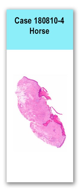
One medium jar of formalin was received. The jar contained two pieces of tissue, the smaller of which was approximately 2 cm x 1.5 cm x 1 cm, soft, pale-tan and overlain with puckered epithelium (urethral process). The larger piece of tissue was 6 cm x 3.5 cm x 1.5 cm and consisted of ulcerated haired skin and underlying soft tissue (ventral penile sheath). On cut section, the tissue was firm, pale-tan, with pinpoint to 0.2 cm diameter well-demarcated white to brown foci.
One section of penile sheath is examined. The dermis is diffusely infiltrated by large numbers of eosinophils, and fewer lymphocytes, plasma cells, macrophages, and neutrophils. Multifocal aggregates of degranulated eosinophils (eosinophilic granulomas) are scattered throughout the dermis, and occasionally, one or multiple profiles of variably mineralized larval nematode are at the center. Larval fragments have a thin, clear to eosinophilic integument and indistinct internal structures with skeletal muscle occasionally discernible. Occasionally, remnant nematodes are mineralized. The epithelium is multifocally ulcerated and a thick crust composed of necrotic eosinophils and epithelial cells overlays these regions. Pale, immature pale basophilic fibrous tissue dissects through dermal collagen bundles.
N/A
Penile sheath: Severe, regionally extensive, chronic eosinophilic and mixed inflammatory posthitis with multifocal superficial ulceration with eosinophilic crust, and multifocal eosinophilic granulomas with intralesional larval nematodes (consistent with Habronema sp.)
The examined sections are consistent with clinical suspicion of Habronemiasis. Eosinophilic infiltrates extend to all borders of the examined sections, and may correlate to extention of this process in surrounding tissues.
Habronemiasis, or “summer sores” commonly occur in the skin of the penis, penile sheath, and around the mouth. It is a hypersensitivity reaction to the parasite, and even after the parasite dies, can continue to cause ongoing inflammation. This is why, oftentimes, owners will pursue surgical excision. Conference goers also discussed how in white-skinned animals, caution should be taken when interpreting these lesions as sometimes skin that is ulcerated by another process such as squamous cell carcinoma will be masked by this inflammatory lesion.N/A.
Case contributor: Hosts- Conference presenters: Drs. Melissa Roy and Kevin Keel