Case 4 180706 (18N1122)
Conference Coordinator: Dr Melissa Roy.
//
Eleven-year-old, female, spayed domestic shorthair cat.
The patient developed progressive respiratory distress over the course of several months. Thoracic radiographs revealed coalescing nodular opacities throughout all lung lobes overlying a diffuse miliary pattern. Bronchioalveolar lavage had suppurative inflammation with marked epithelial cell atypia. Cultures of this fluid had no fungal growth, and only commensal bacterial isolates. The patient was placed on an anti-inflammatory dose of prednisolone based on suspicion for pulmonary carcinoma. Approximately one month later, the patient re-presented with progression of clinical signs, was euthanized, and submitted for necropsy.
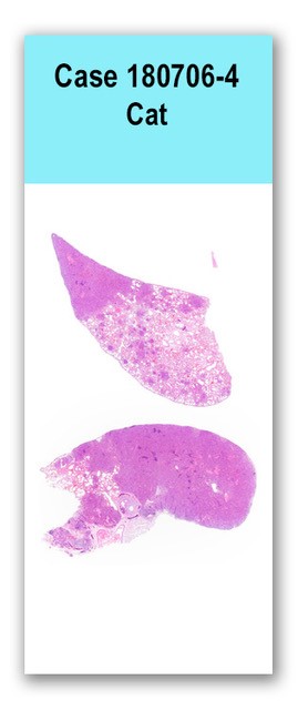
Approximately 80% of the lungs had variably-sized, well-demarcated, pale tan, firm regions of parenchyma. The remaining lungs were soft and mottled pink to red.
Two sections of lung are examined in which 20% to 80% of the parenchyma is infiltrated by a densely cellular, unencapsulated, multinodular neoplasm composed primarily of cuboidal to columnar epithelial monolayers lining alveolar-like structures. Neoplastic cells have distinct cell margins and scant eosinophilic cytoplasm. Nuclei are round with finely stippled chromatin and a single nucleolus. Anistocytosis and anisokaryosis are moderate and there are 15 mitotic figures noted in ten high-powered fields. The mass is infiltrated multifocally by moderate numbers of neutrophils, lymphocytes, and plasma cells. There is frequent single cell necrosis. Up to 25% of neoplastic cells contain 2-14 micron ovoid yeast organisms within the cytoplasm. Organisms are often clustered, and have a thick, non-staining capsule. Bronchi are filled with mucus admixed with necrotic cells. One bronchus appears to be filled with wispy fibrous connective tissue and small caliber vessels (granulation tissue). There are multifocal areas of hemorrhage.
A Gomoris-methenamine silver stain revealed large numbers of intracellular and extracellular, variably-sized organisms with thick capsules.
Ancillary Diagnostic Tests: Fungal culture resulted in the growth of small numbers of Rhodotorula mucilaginosa. A PCR-assay was performed upon the paraffin-embedded tissues, and subsequent sequencing of the related ITS region, confirmed the identity of the fungus.Lungs: Pulmonary carcinoma with intralesional yeast organisms
The lungs have large regions of carcinoma, which based on the histologic appearance, distribution, and lack of identifiable neoplasia anywhere else in the body, is presumed to be a primary pulmonary carcinoma. In addition to this, the neoplastic regions frequently have intracellular variably-sized, GMS-positive yeast organisms. Culture of the fresh frozen tissue revealed small numbers of Rhodotorula mucilaginosa. This was confirmed using PCR. Rhodotorula species are ubiquitous saprophytic environmental yeasts that have previously often been considered nonpathogenic in humans and animals; however recent case reports have implicated Rhodotorula as an emerging pathogen, primarily in immunocompromised patients. Opportunistic infections of skin, meninges, eyes, peritoneum, and lungs have been reported. We thank Dr. Kevin Woolard for his contributions to the histopathology and sequencing.
Wirth F, Goldani LZ. Epidemiology of Rhodotorula: An emerging pathogen. Interdisciplinary perspectives on infectious diseases. 2012; Article ID 465717, 7 pages.