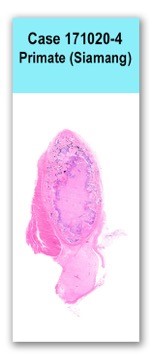
Case 4 171020 (17N2124)
Conference Coordinator: Molly Liepnieks
//
10-year-old, vasectomized male, Siamang
Patient was diagnosed with disseminated coccidioidomycosis at 1 year of age; lesions were present in skin/bone of left hip, right wrist, and in the lungs. The patient did not respond to fluconazole but went into remission on long-term itraconazole therapy. Patient had a four week history of inappetence and lethargy. Previously noted Coccidioides-related lesions were noted to be stable. Ultrasound findings were similar to previous rechecks. IgG titer 1:1 at. Dr Pappaiogianis lab coccidiodes PCR from lung wash negative. Bloodwork was indicative of a hepatopathy and persistent hypoglycemia. Antifungal treatment was halted and liver values improved with supportive care. Liver biopsy was performed. Five days later patient was found dead in his enclosure.
A 10-year-old, male Siamang is necropsied on September 2, 1017 at 9:00 am, more than 24 hours post-mortem. The body is in poor to fair postmortem condition and there are adequate subcutaneous and visceral adipose stores.
The anterior and posterior aspects of the left inferior lung lobe are adhered to the thoracic wall. There are two nodules within the heart, one within each of the ventricles. Each nodule is approximately 2 cm in diameter, tan to red on cut section, and variably mineralized.Heart: On section of heart is examined in which approximately 80% of the section is necrotic with large multifocal to coalescing foci of basophilic to amphophilic, granular material (mineral) and karyorrhectic cellular debris. Small numbers of 60-80 micrometer diameter, round fungal sporangia with a 4-5 micrometer thick, double-contoured, refractile hyaline wall are scattered throughout the necrotic regions. Sporangia are frequently degenerate, but occasionally contain multiple 5-7 micrometer endospores. Cardiomyocytes in the necrotic region are individualized and hypereosinophilic with loss of striation and nuclear detail. Vessels are frequently filled with amphophilic acellular material. Few aggregates of lymphocytes and plasma cells are infiltrating the necrotic region as well the surrounding cardiomyocytes.
No special stains.
Heart: severe, multifocal, chronic granulomatous myocarditis with fibrosis, mineralization, and endosporulating yeast organisms
Lesions noted within the heart are consistent with coccidiodomycosis and are likely the cause of the death. Lesions were extensive and likely contributed to poor cardiac function and potential malignant arrhythmia generation. Endosporulation is indicative of active disease within the heart. Sections of liver and lung did not show evidence of coccidiodomycosis. Due to the risk of zoonotic disease transmission, bones were not examined.
Coccidioides immitis is a dimorphic fungus found in the environment. Infection occurs when arthroconidia, or spores, are inhaled. Infection can become disseminated and spread to bone, spleen, liver, kidneys, heart, eyes, skin, brain, and spinal cord.Jubb, K. V. F., Peter C. Kennedy, and Nigel Palmer. Pathology of Domestic Animals. St. Louis, MO: Elsevier, 2016. Print.

