Case 4 170505
Conference Coordinator: Sebastian Carrasco
//
Six-month-old, female quarter horse
This filly came from a farm at which two other horses reportedly had neurologic disease over past five years. The patient had a grade 4/5 ataxia. Cervical radiographs were unremarkable. She was down in the trailer, and she was unable to stand without assistance upon arrival to the clinic. Neuronal axonodystrophy was suspected. The animal was eventually euthanized due to the poor prognosis.
Four 0.4- to 0.5-cm-diameter ulcers were present on squamous epithelium of the cardia. Hundreds of large robust nematodes consistent with Parascaris equorum filled the duodenum and jejunum. Approximately 50, thin, elongate, white nematodes consistent with strongyle larvae were present in the large intestine, most notably in the right dorsal colon. The aorta at the level of the cranial mesenteric root was segmentally thickened. Other lesions were insignificant and/or were not relevant to the presenting complaints or the slide shown here.
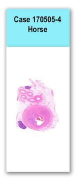
This slide has two sections of small lymph nodes and five sections of muscular arteries, the largest of which has a total diameter of approximately 1.8 cm. The largest artery is asymmetrically thickened. The intima is expanded and partially effaced by multifocal to coalescing necrosis, inflammation and multiple sections of nematodes.. The nematodes are covered by a superficial layer of fibrin with abundant erythrocytes neutrophils and small amounts of cellular debris. The endothelial cells are missing in regions lined bv the thrombus and nematodes. The nematodes are approximately 400 um in diameter with a smooth, glassy eosinophilic, 8.0-um-thick cuticle, platymyarian muscle, and a large central intestine lined by a few multinucleated cells. The reproductive tracts are not visible or are underdeveloped (consistent with larval forms). The adjacent tunica media and adventitia are multifocally vacuolated and distorted with moderate to abundant numbers of infiltrating lymphocytes, plasma cells and neutrophils with multifocal small areas of necrotic cellular debris and some scattered erythrocytes. Tiny aggregates of such leucocytes are scattered throughout the adjoining adipose tissue. The advential vessels of the smaller arteries are cuffed by small numbers of lymphocytes and plasma cells.
No special stains.
Severe, multifocal to coalescing, neutrophilic, lymphoplasmacytic, endarteritis with luminal thrombosis and intralesional larval nematodes (presumed Strongylus vulgaris larvae)
This horse had lesions in the medulla oblongata and thoracolumbar region consistent with equine neuroaxonal dystrophy (NAD), which was likely the cause of the neurological signs seen in this patient. The arterial lesions were incidental to the clinical history, and they were consistent with the migration of S. vulgaris larvae.
Of the strongyles affecting horses, Strongylus vulgaris is by far the most damaging to the host. The lesions seen in this case were caused by migration of the fourth-stage within arteries. This larval migration can eventually lead to tortuous tracts and thrombi in the arterial wall, which may be associated with aneurisms and endarteritis.
The artery was labeled as abdominal aorta, but some conference participants commented on the likelihood that this was the cranial mesenteric artery, based on the size and it’s presence in fatty tissue (possibly mesenteric root) with smaller arteries, and lymph nodes.
The artery was labeled as abdominal aorta, but some conference participants commented on the likelihood that this was the cranial mesenteric artery, based on the size and it’s presence in fatty tissue (possibly mesenteric root) with smaller arteries, and lymph nodes.
Miller LM, VanVleet JF, Gal A. Cardiovascular system and lymphatic vessels. In: McGavin MD, Zachary JF, eds. Pathologic Basis of Veterinary Disease. 5th ed. St. Louis, MO: Mosby Elsevier; 2012;577.
Robinson WF, Robinson NA. Cardiovascular system. In: Maxie MG, ed. Jubb, Kennedy, and Palmer’s Pathology of Domestic Animals. Vol 3. 6th ed. St. Louis, MO: Elsevier; 2016:85-87