Case 3 170505 (CAHFS)
Conference Coordinator: Sebastian Carrasco
//
This is a 2.5-month-old, female Berkshire pig
This pig had a history of fever (103 F), wheezing and inappetence, before dying. Other pigs in the litter exhibited similar signs.
The animal submitted for necropsy was a 49 lb gilt in good body condition with adequate amounts of fat. Multiple lymph nodes were mildly enlarged and red. The right lung lobes were diffusely dark-red and wet. There were no other significant gross lesions.
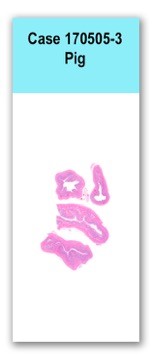
This slide contains four sections of intestine, including ileum and the junction of ileum and colon, in which the mucosal glands are elongated, dilated, branched, and are frequently lined by enterocytes up to eight cell-layers thick (hyperplasia). Some tips of villi tips are necrotic, and are expanded by pale eosinophilic fibrillar material and karyorrhectic cellular debris. The Peyer’s patches are severely depleted and much of the lymphoid tissue is replaced with epithelioid macrophages that are frequently foamy. Crypts are variably expanded by necrotic, eosinophilic cellular debris with small numbers of neutrophils. There is multifocal crypt herniation. Scattered villous and crypt epithelial cells are necrotic. The lamina propria is moderately expanded by plasma cells, lymphocytes, eosinophils, and neutrophils. Ciliated, 30- to 50-micrometer-diameter protozoa (consistent with Balantidium coli) are occasionally scattered throughout the lamina propria and aggregate at the mucosal surface.
A Steiner’s stain revealed numerous comma-shaped bacteria along the apical portions of the affected intestinal epithelium.
Samples of fecal material and/or intestines were negative for rotavirus (ELISA), Salmonella (PCR and culture), transmissible gastroenteritis virus (pcr), swine delta coronavirus (PCR)n and porcine epidemic diarrhea virus (PCR). Samples evaluated for classical swine fever (tonsil) and porcine reproductive and respiratory syndrome virus (lung and spleen) and influenza (lung) were also negative by PCR.
Immunohistochemistry stains for porcine circovirus-2 did not reveal any antigen in the sections of intestine.
Ileum, cecum and colon: Moderate to severe, multifocal to coalescing lymphoplasmacytic, histiocytic, neutrophilic proliferative and necrotizing enterocolitis with Balantidium coli and intracellular Lawsonia-like organisms.
The enterocolitis was the likely cause of death. It was attributed to Lawsonia intracellularis, based on the nature of the lesions and the morphology of the intracellular bacteria. This gram-negative bacteria is an intracellular pathogen that causes disease in a wide range of animals. The disease in pigs occurs in four different forms: porcine intestinal adenopathy, necrotic enteritis, regional proliferative ileitis, and proliferative hemorrhagic enteropathy. Lesions in the ileum and colon in this case were characteristic of regional ileitis and necrotic enteritis. The necrotizing lesions of the intestinal mucosa were interpreted as sequelae of the proliferative enteropathy.
The etiology of the lymphoid depletion in the Peyer’s patches is unknown. Common infectious agents, such as PCV-2 and Salmonella, are commonly associated with lymphoid depletion in pigs, but were ruled out in this case. It is possible that anorexia could have contributed in some degree to lymphoid depletion.
Little is known about the pathogenesis of Lawsonia intracellularis in pigs. This pathogen invades immature enterocytes in the crypts, which causes hyperplasia of the infected cells and leads to proliferative enteropathy. Recent studies have proposed that bacterial replication and dissemination is tied to replication of the enterocyte.
This case was contributed by Dr. Santiago Diab of the California Animal Health and Food Safety System.
Turner JR. The gastrointestinal tract. In: Kumar V, Abbas AK, Fausto N, Aster JC, eds. Robbins and Cotran Pathologic Basis of Disease 8th ed. Philadelphia, PA: Elsevier Saunders; 2009:801.
Guedes RM, Machuca MA, Quiroga MA, Pereira CE, Resende TP, Gebhart CJ. 2017. Lawsonia intracellularis in Pigs: Progression of Lesions and Involvement of Apoptosis. Veterinary Pathology. Mar 24:0300985817698206.
Guedes RM, Gebhart CJ. 2003. Onset and duration of fecal shedding, cell-mediated and humoral immune responses in pigs after challenge with a pathogenic isolate or attenuated vaccine strain of Lawsonia intracellularis. Vet Microbiol. 91:135-145

