Case 2 170707-2 (17B1409)
Conference Coordinator: Wesley Siniard
//
Nine-year-old, male Shubunkin goldfish (Carassius auratus)).
The owner noticed a slowly growing mass surrounding the right eye about one year prior to presentation. In the past month, the mass has begun growing at a faster rate. The fish does not exhibit any clinical signs.
A 2 x 1 x 0.8 cm, soft, multinodular , mottled black to tan to white mass is surrounding an ocular globe.
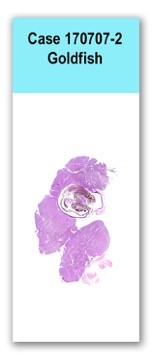
Two parasagittal sections of ocular globe and associated mass are examined. An unencapsulated, densely cellular mass, composed of short interlacing bundles and streams of spindle cells embedded in a fine fibrovascular stroma, surrounds the posterior aspect of the ocular globe and extends to section margins. Neoplastic cells have indistinct cell borders, contain a moderate amount of whispy, amphophilic cytoplasm, which occasionally contains black, finely granular pigment. Th cells have an oval to elongate nucleus with finely stippled chromatin and one to three distinct nucleoli. Anisocytosis and anisokaryosis are moderate and there are four mitotic figures per ten 400x fields. Moderate numbers of melanomacrophages are scattered throughout the neoplastic cell population. Large numbers of lymphocytes are infiltrate the conjunctiva.
The neoplastic cells were negative for Melan-A.
No special stains..
Periocular mass: chromatophoroma
Based on the morphology and the pigmentation of the neoplastic cells, the mass was designated as a chromatophoroma. Melan-A, typically used to identify melanocytic cells, has not been clearly described in non-mammalian species. A negative result does not reliably excludes a melanocytic cell of origin. Chromatophores, include meanophores, iridophores, erythrophores, and xanthophores. The lack of bifringence makes an iridophoroma less likely, but it cannot be ruled out. The only way to definitively diagnose the pigment cell of origin is through electron microscopy. The prognostic significance of the different types of chromatophoromas was discussed amongst conference attendees, however it was decided that the pigment cell of origin is likely not significant for prognosis. The etiology of chromatophoromas is unknown, however it is presumed to be a combination of hereditary, carcinogenic, and age-related factors in fish.
Camus, M, Hyatt, M, Clauss, T, Berliner, A, Camus, A. Chromatophoroma in a crevice kelpfish (Gibbonsia montereyensis). Veterinary Clinical Pathology. 2011; 40/4: 549-552.
Etoh H, Hyodo-Taguchi Y, Aoki K, Murata M, Matsudaira H. Incidence of chromatoblastomas in aging goldfish (Carassius auratus). Journal of the National Cancer Institute. 1983; 70:523-528.
Masahito P, Ishikawa T, Sugano H. Pigment cells and pigment cell tumors in fish. Journal of Investigative Dermatology. 1989; 92: 266S-270S.