Case 3 180629 (18N0385)
Conference Coordinator: Dr Sarah Stevens.
//
Nine-year-old, male, castrated Labrador retriever.
This patient was diagnosed with multicentric B cell lymphoma with ocular involvement approximately eighteen month prior to necropsy. After completing CHOP chemotherapy with subsequent relapse of disease months after, clinical signs progressed and euthanasia was elected.
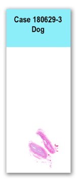
The heart is 0.34 kg (0.59% of body weight) with a left ventricular free wall thickness of 1.7 cm, an interventricular septal thickness of 1.6 cm, and a right ventricular free wall thickness of 0.7 cm. The junctions between the aortic valve cusps are mildly and multifocally hard and thickened (mineralized). Extending off the anterior leaflet of the mitral valve, off the left atrial side of the leaflet, is an irregularly marginated, soft, friable, granular to matte, brown to tan nodule that is approximately 0.5 x 0.4 x 0.4 cm.
A number of peripheral and internal lymph nodes are enlarged including submandibular, superficial cervical, cranial mediastinal and mesenteric root lymph nodes. These lymph nodes range from approximately 1.5 x 1 x 0.8 cm to 3.5 x 3 x 3 cm, with the mesenteric root lymph nodes the least affected and the submandibular lymph nodes the largest. On both superficial and cut surface the lymph nodes bulge, are mottled light tan to pink and are predominantly soft upon palpation.A section of atrioventricular valve has a papillary, proliferative, exophytic mass extending off its surface. The mass is moderately cellular and poorly demarcated. It is composed of short streams and bundles of spindle cells arranged into irregular papillary projections supported by thin stalks of fibrovascular stroma. The cells have scant eosinophilic cytoplasm and an ovoid to elongate nucleus with finely stippled to hyperchromatic chromatin. There is mild anisocytosis and anisokaryosis with 1 mitotic figure identified in 10, 400x, high power fields. The proliferative cells form abundant small capillaries throughout the mass. There is rare to occasional foci of extravasated erythrocytes sometimes associated with clumped bright yellow pigment (hematoidin).
An immunohistochemistry stain demonstrated that the proliferative spindle cells forming the papillary mass were diffusely immunoreactive (membranous to cytoplasmic, stippled) for CD31.
Atrioventricular valve mass: hemangiosarcoma
This small, friable mass was identified unexpectedly extending off the mitral valve, unrelated to the cause of euthanasia. Conference participants were not in agreement regarding the diagnosis in this case. The mass was originally diagnosed as a hemangiosarcoma based on histomorphology and immunohistochemical staining, despite the atypical appearance and location in the body. Other participants discussed the possibility of a non-neoplastic vascular proliferation such as with angiomatosis or a reactive lesion, especially as the mass was not particularly invasive, which might be expected with a malignant neoplasm. Regardless, this valvular mass was not the reason for euthanasia and was considered an incidental finding.