Case 1 180629-1 (18B1205)
Conference Coordinator: Dr Sarah Stevens.
//
Thirteen-year-old, male, castrated mixed-breed cat.
This patient presented to the UCD Soft Tissue Surgery service for removal of a colonic mass. Approximately 1 month prior to surgery, this cat presented to the emergency service for a urethral obstruction, unrelated to the colonic mass. Upon abdominal ultrasound at this visit, a colonic mass was identified and estimated to be approximately 2.5 cm in diameter.
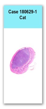
A segment of colon including the mass was removed with 3.5 cm gross margins on either side and submitted for histologic examination.A jar of formalin is submitted containing a segment of luminal tissue consistent with colon. Approximately mid-way through the sample is a concentric, firm, well-demarcated, pale tan mass which expands the wall and extends into and nearly fills the lumen. The mass is approximately 2.5 cm in diameter and is contained within the colon wall.
This slide has a cross section of colon, which has asymmetric mural region that is thickened by an expansile submucosal mass. The mass is well-demarcated, densely cellular, unencapsulated and is supported by a fine preexisting fibrovascular stroma. Cells are arranged in sheets and rows that dissect between preexisting collagen and are round to angular with distinct cell borders, a moderate amount of variably granulated cytoplasm and a round to ovoid, finely stippled nucleus with one to two small basophilic nucleoli. There is minimal anisocytosis and anisokaryosis with rare multinucleated cells and one mitotic figure seen in ten, 400x, high power field. Neoplastic cells abut and compress the inner muscular layer but do not infiltrate into that layer. The mass pushes the mucosal surface into the adjacent lumen, which contains a moderate amount of mucous, occasional hair shafts and granular material.
Neoplastic cells of the colonic submucosal mass have variable numbers of cytoplasmic granules that are highlighted with toluidine blue stain and giemsa stain.
Colon mass: mast cell tumor
Mast cell tumors in cats can be cutaneous or visceral, some of which are associated with mastocytemia. Unlike in dogs, feline cutaneous mast cell tumors are not subdivided into cutaneous and subcutaneous.
Visceral mast cell tumors, such as in this case, are more common in cats than in dogs. According to the literature, eosinophil infiltrates are rare to absent in well-differentiated cat mast cell tumors, in contrast with dogs that consistently have infiltrating eosinophils. However, in pleomorphic or atypical mast cell tumors in cats this is not necessarily true, and increased numbers of infiltrating eosinophils may be seen. Conference participants brought up that, in their experience, feline mast cell tumors tend to be composed of mast cells with minimal to absent granules, which differed from this case where granules were easily identified on routine H&E staining.DJ Meuten. Tumors in Domestic Animals, Ed. 5. 2018. Mast Cell Tumors. Wiley-Blackwell.