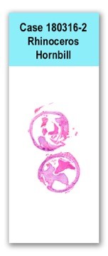
Case 2 180316-2 (18B0362)
Conference Coordinator: Dr Patty Pesavento and Dr Kevin Keel.
//
Five-year-old, male, rhinoceros hornbill (Buceros rhinoceros)
This hornbill was anesthetized for an annual examination and an endotracheal tube was placed. No respiratory abnormalities were identified at that time, and there was no previous medical history related to airway disease.
Two months later a tracheal stricture was diagnosed. The bird was exhibiting raspy breathing during the follow up exam and breathing difficulties persisted. During that time the bird was treated with meloxicam and Enrofloxacin.The bird was anesthetized again two weeks after the diagnosis of a tracheal stricture, because it failed to improve. Radiographs and tracheostomy confirmed that the stricture had worsened and was occluding more of the trachea. A tracheal resection and anastomosis was performed to remove the section of the trachea affected by the proliferative tissue.An approximately 6-cm-long segment of formalin-fixed trachea was received for histopathology. The middle portion of this segment was completely occluded by a mixture of proliferative, slightly gelatinous, tough grey tissue (presumably connective tissue) and yellow friable material. The connective tissue protruded assymetrically from the mucosal surface and filled a approximately fifty percent of the tracheal lumen. The remainder of the luminal space was filled by the yellow, caseous exudate.
This slide includes two transverse sections of trachea. Both are similarly affected by lumenal rafts (plaques) of degenerate heterophils, and asymmetric proliferation of submucosal connective tissue that protrudes into the lumen. A large portion of the caseous exudate contains sheets of closely packed fungal hyphae. The mucosal epithelium adjacent to the thickened connective tissue is hyperplastic. It has numerous goblet cells and occasionally forms large cysts in the submucosa. A portion of the proliferative tissue is covered by squamous epithelium. The mucosal epithelium on the opposite side of the trachea, is variably ulcerated or eroded. A portion of the proliferative tissue is also ulcerated and this segment is adhered to heterophilic exudates which are subtended by a band of multinucleate giant cells. The tracheal lumen contains moderate amounts of mucoid exudate admixed with heterophils, desquamated epithelial cells, dense sheets of fungal hyphae and small numbers of mixed bacteria.
Fungal hyphae have parallel walls and greenish brown fruiting bodies are evident in one focus. Although misshapen, the fruiting bodies consist of vesicles that are often covered by a single row of phialides (consistent with Aspergillus spLarge foci of the tracheal cartilage are necrotic and sheets of degenerate heterophils surrounded by multinucleate giant cells extend through the tracheal wall. A few aggregates of lymphocytes are scattered in the connective tissue adjacent to the trachea.A Gram stain highlighted a mixture of Gram-negative and Gram-positive bacteria of varied shapes. A geimsa-stained section revealed more bacteria than the Gram stain, but they were still not numerous. A Gomori's methenamine silver stain revealed numerous hyphae as previously described. Most of the hyphae and the necrotic cartilage failed to be stained by the periodic acid-Schiff technique.
Severe, chronic, heterophilic to granulomatous, necrotizing and ulcerative tracheitis with intralesional fungal hyphae (consistent with Aspergillus sp) and mixed bacteria
Severe, chronic, focally extensive, luminal, tracheal fibrosis and glandular proliferation (tracheal stenosisPost-intubation tracheal obstruction is a potential complication that is not uncommon in the treatment of birds. In one review nearly 2% of the intubations resulted in obstruction and overall mortality associated with the obstruction was 70%. In the case of nearly complete obstruction, tracheal resection may be helpful. In this case the bird was continuing to improve four weeks after surgery.
Infections are not typically a major confounding factor in tracheal stenosis and it is the obstruction itself which is usually the major morbidity factor. However, the tracheal stenosis in this case may have been the nidus for the Aspergillus infection. A large segment of the proliferative tissue was ulcerated and the manner in which the fungal plaque adhered to the ulcerated surface suggested that interrupted air flow permitted fungal spores to impact upon the mucosal surface and initiate the infection. The large necrotic sequestrum was suggestive of an infarct. The tendency for Aspergillus spp. to invade blood vessels would be consistent with this pathogenesis.Some conference participants commented that the proliferative tissue had the appearance of a polyp. Although it does resemble a polyp in section, this was a segmental change over a broad region of the trachea. However, the proliferation in this case was much less symmetric than many other cases evaluated. Often the proliferative tissue is circumferential.Sykes IV, J.M., Neiffer, D., Terrell, S., Powell, D.M. and Newton, A.. 2013. Review of 23 cases of postintubation tracheal obstructions in birds. Journal of Zoo and Wildlife Medicine, 44(3):700-713.
Clippinger, T.L. and Bennett, R.A. 1998. Successful treatment of a traumatic tracheal stenosis in a goose by surgical resection and anastomosis. Journal of Avian Medicine and Surgery. 12(4):243-247.
