Case 2 171208-2 (17B2637)
Conference Coordinator: Devinn Sinnott.
//
10-year-old, FS, Labrador retriever.
During a routine dental cleaning by a referring veterinarian, a mass was noted on the ventral aspect of the neck. Her T4 value at this time was 6.3 ug/dl, and her owner reported increased panting, appetite, and water consumption, weight loss, and hair coat changes. Blood work and chest radiographs performed at a referral specialist hospital were normal, and fine needle aspirate of the mass showed epithelial cells consistent with a thyroid carcinoma. She was referred to UCD Oncology, where a 4 cm, freely movable left thyroid mass was palpated and confirmed by ultrasound. A thyroidectomy was perfomed to remove the mass.
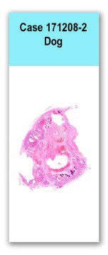
A 5.1 x 3.6 x 2.5 cm, multinodular, smooth, mottled pale pink to dark red, firm mass was received. On cut section the mass had multiple cavitations ranging in diameter from 3-11 mm that contained thin, clear, red fluid. The mass also contained anastomosing trabeculae of white, hard material.
Examined is one section of a large, well demarcated, encapsulated mass composed of cells arranged in islands of disorganized follicles and sheets separated by thick bands of dissecting fibrous tissue. The cells are polygonal with variably distinct cell borders, abundant eosinophilic cytoplasm, and a single, round, central nucleus with finely stippled chromatin and 1-3 basophilic nucleoli. Anisocytosis is mild, and anisokaryosis is moderate. No mitoses are seen in ten 400x fields. Follicles frequently contain homogeneous, eosinophilic material that is occasionally mineralized. The center of the mass has multiple large areas of necrosis and hemorrhage. Multiple islands of anastomosing trabeculae of woven bone and chondroblasts embedded in a basophilic, chondroid matrix surrounded by a thick band of fibrous tissue are scattered throughout the mass.
No special stains.
Thyroid gland: follicular to solid thyroid carcinoma with osseous and chondroid metaplasia
Thyroid carcinomas occur more commonly in dogs (90%) than thyroid adenomas (9%). There is no sex predilection for thyroid carcinomas in dogs, and Boxers, beagles, Siberian huskies, and golden retrievers have a higher incidence compared to other breeds (1). Unlike cats, most dogs with functional thyroid neoplasms are euthyroid or have mildly elevated T4/T3 values. Clinical signs in dogs may include polyuria, polydipsia, weight loss despite increased appetite, muscle weakness, heat intolerance, and nervousness. Large, invasive tumors may cause dysphagia or dyspnea (1).
Histologically, thyroid carcinomas are classified as follicular, solid/compact, or papillary based on the predominant pattern of growth (1). In follicular thyroid carcinomas, neoplastic cells form follicular structures that often contain clumped and mineralized colloid (1). Solid or compact thyroid carcinomas form solid sheets of neoplastic cells separated by fibrous stroma, and immunohistochemistry for thyroglobulin, thyroid transcription factor 1, or calcitonin may be necessary to distinguish these tumors from C-cell carcinomas (1). Follicular to solid type carcinomas are the most common subtype in dogs. Papillary thyroid carcinomas form papillary projections that extend into cystic spaces within the mass, and are rare in domestic species compared to humans (1). Undifferentiated thyroid carcinomas, including small cell and giant cell variants, are uncommon in domestic animals (1). Canine thyroid carcinomas are often partially encapsulated and contain large central areas of necrosis and hemorrhage. Areas of mineralization or bone formation can also occur within the mass (1). Osseous and chondroid metaplasia of the stroma has been reported in canine thyroid carcinomas (2, 3). Malignant mixed thyroid tumors can also occur, in which both the follicular cells and mesenchymal components of the mass are malignant. The mesenchymal components are often osteogenic or cartilaginous, and may appear similar to osteosarcoma or chondrosarcoma (1, 4). Thyroid carcinomas eventually breach their capsule and invade into local structures including the trachea, esophagus, and larynx. Invasion into the thyroid or jugular veins creates neoplastic thrombi, which often leads to metastasis to the lungs (1).1. Meuten DJ, editor. Tumors of domestic animals, 5th edition. Ames, IA: John Wiley & Sons, Inc.; 2017. p. 791-799.
2. Kobayashi R, Yamada N, Kitamori T, Kitamori F, Sato K, Doi T, Wako Y, Sato J, Tsuchitani M. Follicular thyroid carcinoma characterized by adundant stromal components with chondroid and osseous metaplasia in a dog. Journal of Veterinary Medical Science. 2014. 76(8): 1161-1164. 3. Brodey RS, Kelly DF. Thyroid neoplasms in the dog: a clinicopathologic study of fifty-seven cases. Cancer. 1968. 22(2): 406-416. 4. Grubor B, Haynes JS. Thyroid carcinosarcoma in a dog. Veterinary Pathology. 2005. 42: 84-87.