Case 2 170526-2 (11N1963)
Conference Coordinator: Sarah Stevens
//
The subject was a 2-year-old, male, African pygmy hedgehog (Ateletrix albiventris.
This hedgehog had a two-month-long history of respiratory disease and ataxia, before he was initially presented to a veterinarian for stumbling in his cage. At that time, he was mildly ataxic without appreciated conscious proprioceptive deficits and no cranial nerve deficits. Two months after the initial visit, he was readmitted for self-traumatization of his right, rear leg followed by listlessness. The clinical signs were suggestive of wobbly hedgehog syndrome. Euthanasia was elected because of the progressive clinical signs and the apparently poor prognosis.
The only significant gross lesions were erosion and ulceration of the skin, particularly around the digits. This was thought to be consistent with the history of self-induced trauma.
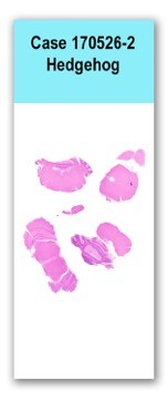
This slide has several coronal sections through the brain, including a section at the level of thalamus and hippocampus, a section at the level of the pons, and two sections including cerebellum and medulla. Most sections have bilateral regions of vacuolization, but these changes are most severe in the brainstem and the cerebellar white matter. The affected areas also have multifocal, moderate swelling of myelin sheaths, mild gliosis and rarely macrophages within myelin sheaths (digestion chambers). There is also a moderate number of shrunken and hypereosinophilic neurons consistent with neuronal necrosis.
A luxol-fast blue stain indicated a loss of myelin in regions of vacuolization.
Brain: Severe, multifocal, white-matter vacuolation and demyelination with mild to moderate neuronal necrosis, mild gliosis and rare axonophagia (consistent with wobbly hedgehog syndrome).
The clinical signs and histologic changes in this animal are consistent with a neurologic disease of captive-bred African hedgehogs (Atelerix albiventris) that has come to be known as wobbly hedgehog syndrome. The disease is not known to occur in other species or in wild populations, and it is hypothesized to be a genetic disease, possibly associated with limited genetic diversity in the captive populations. The pathogenesis is not understood, however, several publications describe this condition and the associated histologic changes. Vacuolation and demyelination tend to predominate in the white matter of the cerebrum, cerebellum and brainstem, as well as within the spinal cord. This is thought to primarily represent demyelination with secondary neuronal degeneration and necrosis. Inflammation is not reported as a feature of this disease.
Some pathologists reviewing the case thought that the self-induced trauma was a little bit unusual. The comment was made that this disease typically affects motor tracts and spares the sensory tracts. Therefore, the stimulus for the trauma was uncertain.
Graesser D, Spraker T, Garner M, Madri J, et al. 2006. Wobbly hedgehog syndrome in African pygmy hedgehogs (Ateletrix spp.). Journal of Exotic Pet Medicine. 15(1):59-65.