Case 2 170505-2 (CNPRC Case)
Conference Coordinator: Sebastian Carrasco
//
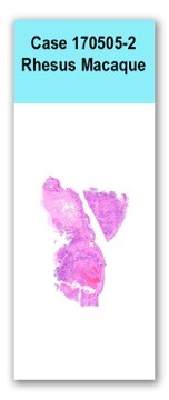
Placenta and aborted fetus from a rhesus macaque at 162 days of gestation.
This fetus comes from a multiparous macaque that is housed outdoors at a primate center. The dam previously had three uneventful pregnancies.
The specimen submitted for necropsy was a full-term fetus with the placenta. Internal organs and brain were severely autolyzed. The maternal surface of the placenta had nine, light-tan, semi-firm, slightly raised nodules. On cut section, these nodules were tan to yellow, dry and semi-firm.
Two sections of placenta are examined in which 30% to 60% of the tissue is necrotic, characterized by regions of increased eosinophilia, loss of cellular detail, and the presence of streaming cellular debris. Multifocal basophilic concretions (dystrophic mineralization) are present in the vicinity of the trophoblasts. The villi are expanded by large numbers of necrotic and viable neutrophils, with fewer numbers of lymphocytes and plasma cells. There are large areas of hemorrhage and fibrin. Stippled basophilic material is abundant throughout the placenta. Occasional clumps of golden brown pigment (presumably hemosiderin) are also present. The decidualized stroma and trophoblastic shell are infiltrated by large numbers of neutrophils with small numbers of lymphocytes and plasma cells. Vessels are frequently lined by hypertrophied, reactive endothelial cells, and small numbers of neutrophils are present within vessel walls (transmigration).
Large numbers of Listeria monocytogenes were cultured from the placenta.
Immunohistochemistry for Listeria spp. indicated clusters of Listeria antigen in regions necrosuppurative inflammation
Placenta: Severe, multifocal to coalescing, necrotizing and suppurative placentitis
The necrosis and Neutrophilic inflammation affecting chorionic villi and intervillous spaces is consistent with Listeria infection. Listeria monocytogenes is a facultative intracellular, gram-negative bacteria that replicates inside phagocytes and can also invade epithelial cells. The pregnancy-associated form of listeriosis causes stillbirths, abortion, and premature birth, and is the most common problem seen in rhesus macaques, at the facility, which housed this dam. The bacteria can cross the placental barrier and hematogenously infect the fetus. Lesions in fetus are not commonly seen as they are often in an advanced stage of autolysis at the time of abortion. Listeria monocytogenes is a ubiquitous pathogen and that can persist in the environment (e.g. water and soil). For this reason, attendees discussed the possibility that nonhuman primates may be getting exposed to L. monocytogenes by ingestion of contaminated food or water.
We offer a special thanks to Dr. Ross Tarara for his contributions to the review of the case materials.
Lemoy MJ, Lopes DA, Reader JR, Westworth DR, Tarara RP. 2012. Meningoencephalitis due to Listeria monocytogenes in a pregnant rhesus macaque (Macaca mulatta). Comparative Medicine. 62(5):443-7.
Wolfe B, Wiepz GJ, Schotzko M, Bondarenko GI, Durning M, Simmons HA, Mejia A, Faith NG, Sampene E, Suresh M, Kathariou S. 2017. Acute fetal demise with first trimester maternal infection resulting from listeria monocytogenes in a nonhuman primate model. mBio. 8(1):e01938-16.
Lecuit M. 2005. Understanding how Listeria monocytogenes targets and crosses host barriers. Clinical Microbiology and Infection. 11(6):430-6.