Case 1 170707-1 (17N1171)
Conference Coordinator: Wesley Siniard
//
10-year-old, male castrated golden retriever
This dog was presented to UC Davis after a three month history of intermittent pallor, a ten day history of tachypnea and lethargy, and a 24 hour history of progressive coughing and dyspnea. The referring veterinarian identified a 3-cm-diameter, heterogenous splenic mass on abdominal ultrasound and a severe nodular pulmonary pattern on thoracic radiographs. The clinical suspicion was that the patient had splenic hemangiosarcoma which had metastasized to the lungs. Due to concern for quality of life and the limited treatment options available, he was euthanized.
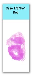
One hundred to one hundred and fifty, multifocal to coalescing, firm, pale tan nodules ranging from 0.3 to 2.5-cm-diameter were scattered throughout all lung lobes, affecting approximately 85 to 90% of the lungs. A large, 7 x 4.5 x 2 cm, similar mass was also noted on the left side of the trachea just caudal to the epiglottis. Twelve nodules ranging from 0.5 to 2.5-cm-diameter were multifocally throughout the spleen and were elevating the capsule and extending into the parenchyma. On cut surface, these nodules were similar to the surrounding splenic parenchyma.
This slide has one section of lung, in which approximately 70% of the normal parenchyma is replaced by three well-demarcated lobules of a densely cellular neoplasm that compress the surrounding tissue. The neoplasm consists of solidly cellular sheets or clusters of rosette-like acinar structures, separated into nests or packets by thin bands of a fibrovascular stroma. Tumor cells tend to have basilar nuclei that palisade against the surrounding stroma, and acinar structures are either empty or contain small amounts of eosinophilic to pale basophilic material, and sometimes small amounts of necrotic cellular debris. The cells have small amounts of granular or flocculent, eosinophilic cytoplasm and poorly defined cytoplasmic borders. Nuclei are round with finely stippled chromatin and variably distinct nucleoli. Anisocytosis and anisokaryosis are mild to moderate, and there are 33 mitotic figures in ten 400x fields. Scattered individual cells throughout the tumor have hypereosinophilic cytoplasm and pyknotic nuclear material. The larger lobules are surrounded by thin but dense bands of fibrous connective tissue populated by plump fibroblasts, often with prominent small-caliber vasculature. In the parenchyma surrounding tumor lobules, alveoli contain large numbers of macrophages with variably foamy cytoplasm, admixed with fewer lymphocytes and plasma cells.
No special stains.
Lungs: metastatic adenocarcinoma (presumed)
Tumors within the lungs are presumed to be adenocarcinomas based on morphology, however the site of the primary tumor was not definitively identified. The cervical mass noted grossly was also completely infiltrated with this neoplasm. This may be the primary site of neoplasia or may be an additional metastasis. Differentials include thyroid carcinoma, parathyroid carcinoma, and anal sac gland adenocarcinoma. Although both lobes of the thyroid gland were identified grossly, this tumor could have arisen from ectopic thyroid tissue.
The splenic masses were consistent with nodular hyperplasia histologically; however, a few suspicious cells were scattered throughout the grossly normal splenic parenchyma. Immunohistochemistry to detect hemangiosarcoma was performed (CD31), and weakly labeled few of these cells. For this reason, a presumptive diagnosis of splenic hemangiosarcoma was made.